The ear is a complicated structure with theee main parts. The external part consists of the pinna and the ear canal. The middle ear consists of the ear drum and an air filled space behind the ear drum. This contains the hearing bones. The air in the middle ear is exchanged via a tube that connects it with the back of the nose. This is called the Eustachian tube. The inner ear is where the actual organ of hearing is situated. This is called the cochlea. It also contains the balance organ or vestibular apparatus. The cochlea and balance organ are connected to the brain by a nerve that allows you to interpret sound and detect changes in your balance. The diagram below shows the different parts of the ear.

The picture below shows the appearance of a normal ear drum. The membrane is thin enough to see the shadows of the structures behind the ear drum. If you move the cursor over the picture some of the main structures become highlighted.

Symptoms of Ear Problems
There are only 5 symptoms that can arise as a result of problems with the ear. These are:
- Hearing loss
- Earache
- Discharge
- Tinnitus
- Imbalance
Your description of the symptoms that you experience will help your doctor make a diagnosis.
Examination of the Ear
The ear can usually be easily examined using an instrument called an otoscope. This is a battery driven special torch designed to be placed in the ear canal. The picture below shows an ear being examined with an otoscope.

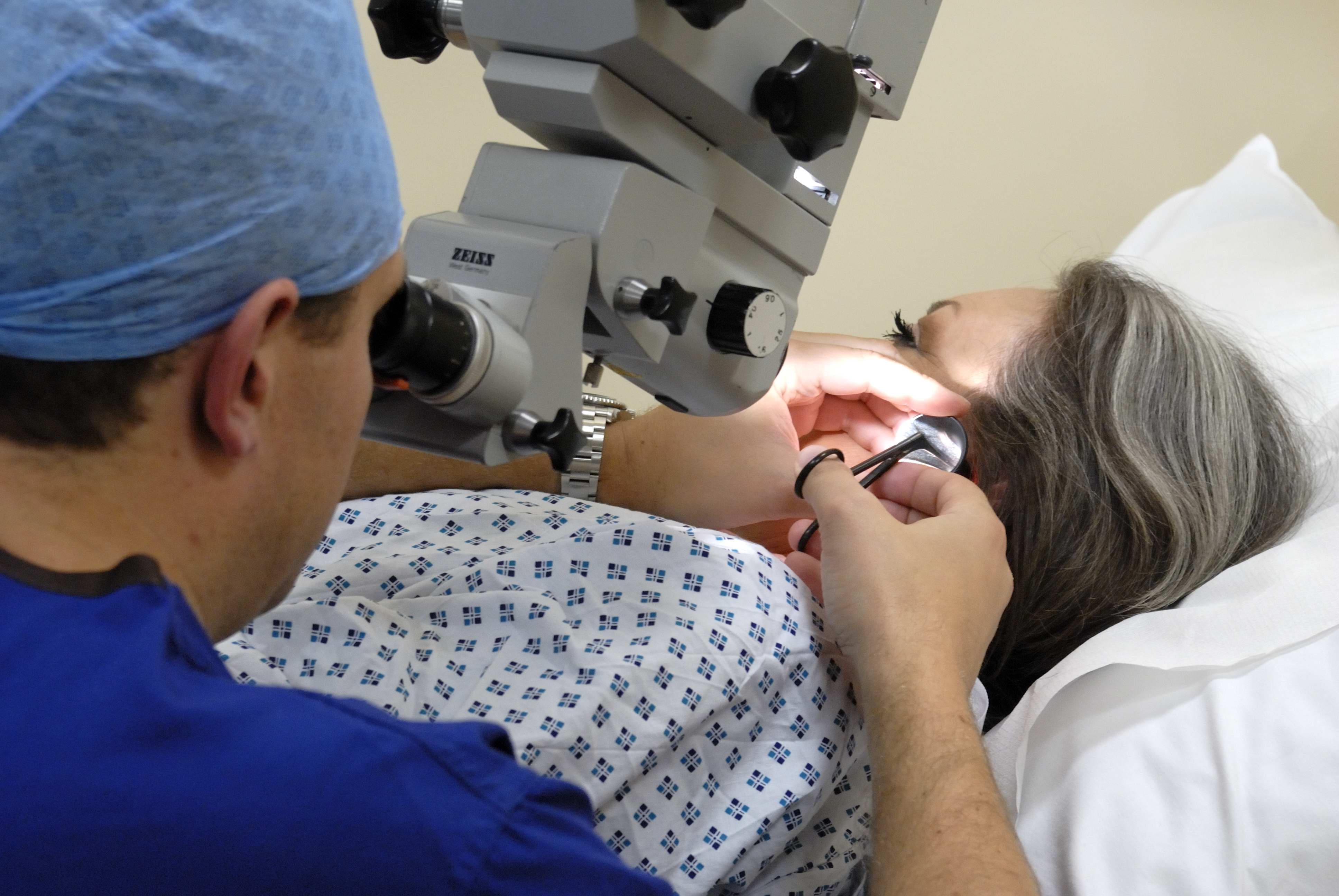
An ear specialist often also uses a microscope to examine the ear drum and ear canal more carefully. The extra magnification that the microscope offers makes making the correct diagnosis much easier. An ear being examined using a microscope is shown below.
As well as having your ear examined, anyone with significant ear symptoms will have a full examination of the balance system. In some situations, it is also necessary to perform a full neurological examination of the head. The examinations required will be explained before they are performed.
Investigation of Ear Problems
In many patients with ear problems it is important to check the hearing. This check is called an audiogram and is performed by an Audiologist. The test involves playing tones into the ear at different frequencies (measured in kilohertz) and dropping the loudness of the sound down (measured in decibels abbreviated to dB) to a level at which it can only just be heard. This is called the hearing threshold. This process is repeated over a number of different frequencies and the thresholds are plotted on a graph. A typical normal audiogram is shown below. In this case the hearing thresholds are all better than 20dB which is the cut off for normality. The thresholds in the right ear are marked in red and the thresholds for the left ear are marked in blue.

If there is hearing loss then the thresholds will be below 20dB. The picture below shows the loudness and frequency of some every day sounds.

Sometimes it is also necessary to perform other types of test after your initial consultation. This might include some more detailed balance function tests or scans which might be either a magnetic resonance imaging (MRI) scan or a computed tomography (CT) scan. You will be told what to expect if you need to have these investigations.
All three parts of the ear can develop problems. The vast majority of problems relate to infection or inflammation of the ear.
The Discharging Ear
When an ear starts to discharge it usually means that there is an infection present. The infection usually affects the middle ear but it can sometimes happen when there is an outer ear infection. Outer ear infections, however, more commonly cause pain and are therefore dealt with in more detail under the heading ‘Earache’. In this section the focus is on infections of the middle ear.
There are two important types of middle ear infection (also called otitis media). The commonest is called acute otitis media. The other type is called chronic otitis media. The main difference from a symptom point of view is the length of time the symptoms have been present. Acute otitis media usually lasts a few days. Chronic otitis media lasts longer than 3 months. The underlying cause of the two types is different.
Acute otitis media
Acute otitis media is the usual cause of ear infections in children although it can affect adults. It is usually a viral infection but can be bacterial. There is usually a high temperature and you feel generally unwell. There is usually earache on one side. There can also be discharge from the ear. Young children can not tell you that they are in pain. They usually pull at the ear instead. The flow chart below shows how acute otitis media develops, click on the picture to enlarge.

Chronic otitis media
- Due to a perforated ear drum
- Due to cholesteatoma
How to Tell the Difference Between Types of Ear Infection
It is often possible to tell which type of ear infection has occurred from the symptoms and it is almost always possible to make a diagnosis by looking in the ear with an otoscope. The table below shows the common features of the different types of ear infection, click on the picture to view an enlarged version:
